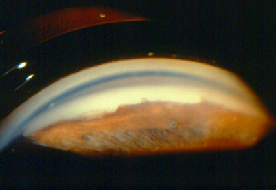
Glaucoma is a difficult disease to detect without regular, complete eye examination, since it usually has no symptoms. To help determine if the drainage channels for the aqueous humour within your eye are open, your eyecare provider will perform a gonioscopy.
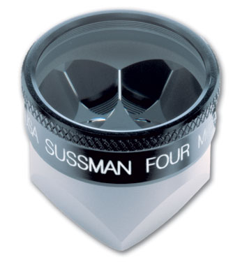
 During gonioscopy, your ophthalmologist will ask you to sit in a chair facing the microscope (slit lamp) used to look inside your eye. You will place your chin on a chin rest and your forehead against a support bar while looking straight ahead. After your eye has been numbed with eyedrops, your ophthalmologist gently places the goniolens on the front of your eye and directs a narrow beam of light into your eye while looking through the slit lamp to examine the drainage angle. (One type of goniolens, the Sussman Four Mirror lens, is show.)
 Determining if the drainage angle of the eye is closed or nearly closed helps your ophthalmologist determine which type of glaucoma you have. Gonioscopy can also detect scarring or other damage to the drainage angle that may explain the cause of certain types of glaucoma. By applying gentle pressure on the lens, the iris is pushed backward to reveal any scarring (synechiae) adhering the peripheral iris to the angle structures.
If your ophthalmologist finds that the drainage angle is closed, a special laser (peripheral iridotomy) can make a small opening in the iris to open the angle. Laser treatment to open the drainage angle may decrease the pressure in the eye and help control glaucoma.
(c) 2009 Robert M. Schertzer, MD, MEd, FRCSC based on 2007 The American Academy of Ophthalmology
Categorized in: Glaucoma
This post was written by Rob Schertzer