
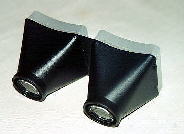
 Photographic images of the optic disc are considered the standard for baseline documentation of the appearance (form) of the optic nerve. However, since the advent of such 3-D imaging devices as the Heidelberg Retinal Tomogram and other nerve fiber layer analyzers, they are often supplanted by these newer modalities. That being said, there is something about the timeless quality of a stereo pair of 35mm photographic slides, seen through a rudimentary stereo viewer, that always makes them useful to have if available.
Glaucoma damage is seen clinically as loss of the nerve fiber layer and an associated thinning of tissue at the optic nerve head. With this damage, ophthalmologists (Eye M.D.s) look for what they call “cupping” of the optic nerve. Stereoscopic disc photos of the optic nerve are helpful in providing a baseline of information about the optic nerve’s condition for future comparison. These photographs are taken in the ophthalmologist’s office using a special camera that can create a stereo image.
Because one ophthalmologist may interpret the appearance of optic nerve cupping differently from another ophthalmologist, optic disc photography is invaluable because it helps create a baseline for future comparison. Your ophthalmologist later may take additional pictures for side-by-side comparison. These can help identify signs of glaucoma progression.
Despite many new imaging techniques for glaucoma, disc photos and a careful clinical examination are still the standard of care for glaucoma.
(c) 2009 Robert M. Schertzer, MD, MEd, FRCSC based on 2007 The American Academy of Ophthalmology