Structural and functional tests
In addition to the function assessed by tests such as Visual Fields, Visual Acuity, and colour vision, an essential ancillary test for glaucoma patients or suspects is 3D imaging of the optic nerve, the surrounding nerve fibre layer, and, more importantly, the ganglion cell thickness in the macular area. These structural tests are correlated with the functional test results to look for progression of glaucoma.
Ocular Coherence Tomography is now the standard
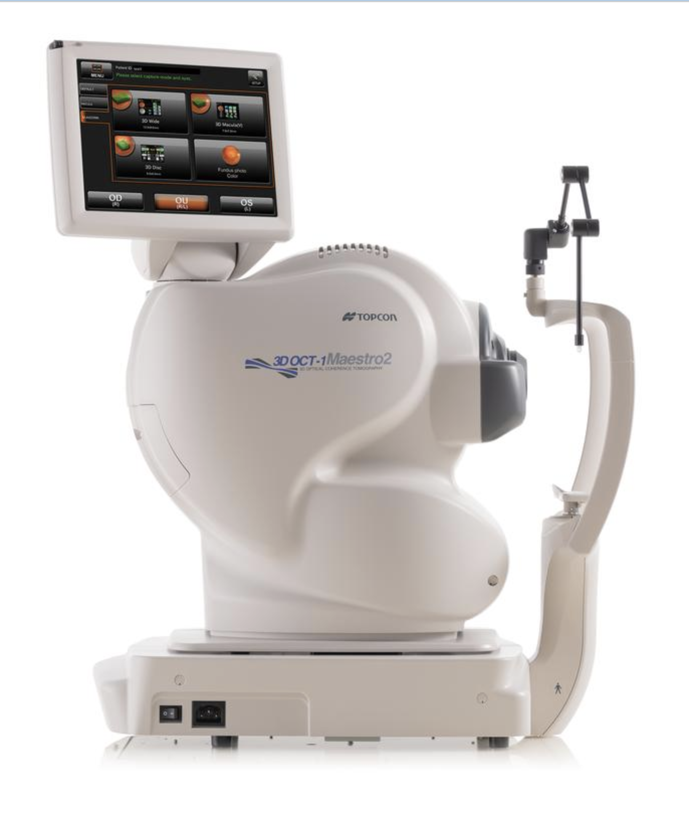 In our practice, the Heidelberg Retinal Tomogram (HRT) is no longer being used for 3D imaging of the optic nerve. It had a proven track record from 1994 to 2012. The Ocular Coherence Topography devices (OCT) have since replaced the HRT as the most effective method for detecting subtle structural changes associated with glaucoma, often before any functional changes are evident on Visual Field testing.
Early in the disease process of glaucoma, individual nerve fibres in the eye’s optic nerve are lost, causing an associated pattern of nerve-fibre-layer thinning. This problem can later translate into loss of tissue at the optic nerve head, resulting in visual field defects and, ultimately, loss of vision.
Both tests are done in the ophthalmologist’s office. During these tests, the patient is required only to remain still while the image is scanned.
The invention of the OCT
© 2025, 2020 Dr. Robert Schertzer Inc.