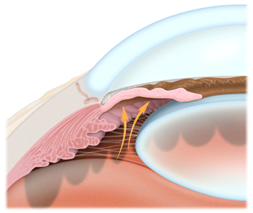
If your ophthalmologist (Eye M.D.) suspects that you have “narrow” or “closed” angles, this means that the drainage channel of your eye is blocked or nearly blocked, placing you at high risk for elevated intraocular pressure and vision loss. This is called angle-closure glaucoma.

An acute attack of angle-closure glaucoma is marked by very high eye pressure and complete blockage of the drainage channel in the eye. Symptoms include pain, red eye, and decreased vision.
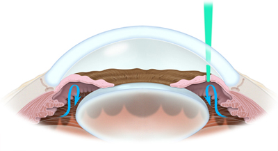
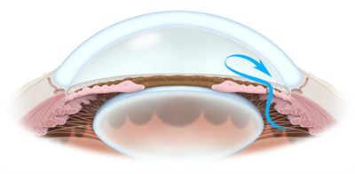
To treat angle-closure glaucoma, your ophthalmologist will perform a peripheral iridotomy (PI), creating a surgical opening within the upper part of the iris (the colored part of the eye) using a laser. This opening is typically so small that it cannot be seen with the naked eye. The opening in the iris allows fluid to flow from behind the iris through the opening, allowing the iris to fall back into a more normal position and opening the drain.


This laser treatment is always performed on an outpatient basis, often in the ophthalmologist’s office. The treatment will not improve your vision, but it can help prevent vision loss from a dangerous type of glaucoma. The side effects of the treatment can include the appearance of a “light streak,” a temporary rise in intraocular pressure, and inflammation. Prior to the laser treatment, you may receive a drop of pilocarpine to constrict your pupil; this can cause a headache around your eyebrow. After the treatment, you will receive a prescription for an anti-inflammatory drop such as prednisolone acetate to use for a few days to prevent inflammation. Follow up visits will be arranged to check for inflammation, make sure the iridotomy is patent and that the angle has widened.
(c) 2015, 2025 Robert M. Schertzer, MD, MEd, FRCSC based on 2007 The American Academy of Ophthalmology


